What Blood Tests Are Used to Diagnose Amyloidosis?
Table of Contents

Amyloidosis is a rare, complex disease caused by the deposition of misfolded proteins—known as amyloid—in various organs and tissues. Early and accurate diagnosis is crucial for managing the disease, improving outcomes, and tailoring treatment approaches. One of the key steps in diagnosing amyloidosis is blood testing, which provides critical insights into the presence, type, and extent of amyloid protein abnormalities.
In this detailed article, we explore the blood tests used in diagnosing amyloidosis, their role in differentiating its types, and how they contribute to a complete diagnostic picture.
1. Introduction to Amyloidosis
Amyloidosis is a group of disorders characterized by abnormal deposition of amyloid fibrils—misfolded protein aggregates—in tissues. Depending on the protein involved, different types of amyloidosis exist, such as:
- AL (Light-chain) Amyloidosis
- AA (Secondary) Amyloidosis
- ATTR (Transthyretin) Amyloidosis – Hereditary or Wild-type
Each has different etiologies, clinical presentation, and detection marker—especially in blood and urine.
2. Why Blood Tests Matter
Blood tests are typically the first choice in diagnosing suspected amyloidosis. They:
Detect abnormal proteins or markers
Identify organs involved
Narrow the type of amyloidosis involved
Guide decisions on biopsy and imaging
Monitor progression of disease and response to treatment
3. Overview of Blood Tests for Amyloidosis
Key blood tests for amyloidosis include:
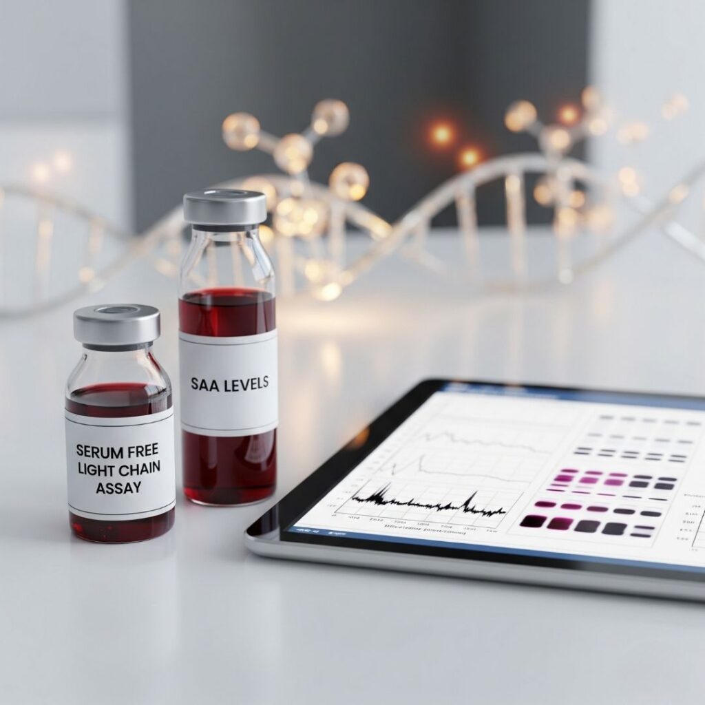
- Serum Free Light Chain (FLC) assay
- Serum Amyloid A (SAA) level
- SPEP and IFE
- Genetic testing for hereditary forms
- Beta-2 microglobulin levels
- Troponin and NT-proBNP for cardiac involvement (supportive)
Let’s explore each in depth.
4. Serum Free Light Chain (FLC) Assay
What it Measures:
Quantifies free kappa and lambda light chains not bound to immunoglobulins.
Why It’s Important:
- Abnormal ratio or increased level is a good indicator of AL amyloidosis
- Useful in identifying plasma cell dyscrasia
- Critical for monitoring treatment response
Normal Ranges:
- Kappa: 3.3–19.4 mg/L
- Lambda: 5.7–26.3 mg/L
- Kappa/Lambda ratio: 0.26–1.65 (outside this indicates monoclonal process)
FLC testing is very sensitive and can identify abnormalities that are not detected by SPEP/IFE.
5. Serum Amyloid A (SAA) Levels
What it Measures:
SAA is an acute-phase reactant protein, mainly raised in AA amyloidosis.
Role in Diagnosis:
- Increased in chronic inflammation and autoimmune diseases
- SAA overproduction is associated with AA amyloid deposition
- Monitoring SAA levels is helpful in evaluating activity of disease
Diagnostic Value:
High SAA + chronic inflammatory disease = possible AA amyloidosis
trend of response to treatment is assessed by fall in SAA levels
6. Serum and Urine Protein Electrophoresis (SPEP/UPEP)
Rationale:
Identifies monoclonal proteins (M-protein) which are commonly seen in association with AL amyloidosis.
How It Works:
- Separates serum proteins by charge and size
Identifies abnormal “M-spike” in the gamma region
Limitations:
- May miss low-level light chain-only diseases (why FLC testing is so vital)
Used with immunofixation for higher precision
Urine testing often involves 24-hour collection to detect the presence of Bence-Jones proteins (light chains).
7. Immunofixation Electrophoresis (IFE)
Purpose:
Identifies the precise type of monoclonal protein present in SPEP or UPEP.
Significance in Amyloidosis:
- Distinguishes IgG, IgA, IgM, kappa, or lambda light chains
- Assists in confirming diagnosis of AL amyloidosis
IFE is more sensitive than SPEP and frequently used in combination.
8. Genetic Testing for Hereditary Amyloidosis
Indications:
- Suspected ATTR amyloidosis with a family history
- Patients with early-onset cardiomyopathy or neuropathy
What is Tested:
- TTR gene mutations (e.g., Val30Met, Thr60Ala)
- Other uncommon amyloid-associated mutations (e.g., ApoA1, ApoA2)
Benefits:
- Confirms ATTRm (hereditary) vs ATTRwt (wild-type)
- Assists in family screening and counseling
9. Beta-2 Microglobulin Testing
A marker of:
- Plasma cell activity
- Kidney function
- Disease burden in AL amyloidosis and multiple myeloma
Interpretation:
- Increased levels in AL amyloidosis
- Reflects renal impairment and prognosis
10. Other Biomarkers in Amyloidosis
Additional Blood Tests:
- Troponin T/I and NT-proBNP: Assess cardiac involvement (particularly in AL/ATTR)
- CRP/ESR: Inflammatory markers
- Liver enzymes: For hepatic involvement
- Creatinine and urea: Indicators of renal function
These form a multi-organ assessment profile.
11. How Blood Tests Help in Subtyping
To ensure accurate diagnosis, it is essential to know the type of amyloid protein, for which blood tests can provide direction:
| Type | Key Blood Test Markers |
|---|---|
| AL | Abnormal FLC, M-spike, IFE-positive |
| AA | Elevated SAA, CRP/ESR |
| ATTR | Negative FLC, genetic mutation in TTR gene |
Subtyping is vital because treatment is radically different among types.
12. Role in Monitoring and Prognosis
Following diagnosis, repeated blood tests assist:
- Monitoring for response to treatment
- Identifying relapse
- Predicting survival
Examples:
- Reduction in FLC levels = effective treatment
- Stable/rising NT-proBNP = continued cardiac strain
- Sustained elevated SAA = lack of control of inflammation in AA amyloidosis
13. Challenges and Limitations of Blood Tests
Challenges Include:
- Small amounts of light chains may go undetected
- M-protein-negative AL amyloidosis is uncommon but exists
- Kidney dysfunction leading to misinterpretation of FLC ratio
- Not all laboratories are equipped with genetic or sophisticated testing
Therefore, blood work needs to be supplemented with biopsy and imaging to get the whole picture.
14. Real-World Case Examples
Case 1: Concealed AL Amyloidosis
A 60-year-old patient presented with fatigue and renal impairment. SPEP was normal, but FLC was positive for increased lambda light chains. Kidney biopsy established AL amyloidosis.
Moral: FLC work can uncover what SPEP does not.”.
Case 2: Incorrect diagnosis of ATTR Amyloidosis
A 75-year-old patient with heart failure symptoms. Blood tests showed normal FLC, negative IFE but genetic testing showed she has hereditary ATTR mutation.
Takeaway: ATTR often does not have abnormal blood markers-genetic testing is very important.
15. Frequently Asked Questions
Q1: Can blood tests diagnose amyloidosis in the blood?
A: No. Blood tests indicate a potential but biopsy with Congo red staining confirm it.
Q2: Why is the free light chain test so important?
A: It identifies light chain abnormalities in AL amyloidosis even in cases with a normal SPEP.
Q3: What if all the blood tests are normal?
A: ATTR or AA amyloidosis can be unassociated with blood abnormalities; imaging, biopsy, or genetic studies are required.
16. Conclusion
Blood tests have an important role to play in the diagnosis and treatment of amyloidosis, particularly in identifying abnormal proteins, dividing the disease into subtypes, and monitoring therapy. But they are not solo diagnoses—biopsies and other sophisticated tests must follow.
It is in the best interest of timely diagnosis and better patient outcomes that one understands which tests to order—and how to interpret them. When there’s an amyloidosis suspicion, prompt evaluation using the appropriate combination of blood tests, imaging, biopsy, and genetic analysis is the forward-looking approach.

