
Restrictive Cardiomyopathy Explained:
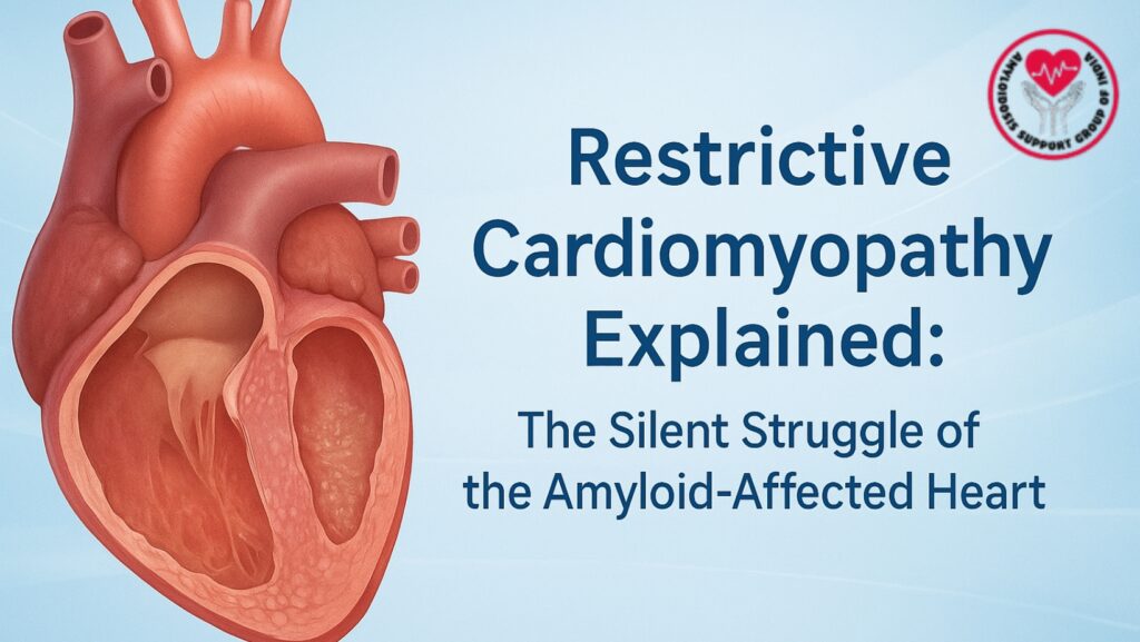
Table of Contents
Introduction
The human heart is an amazing organ. It tirelessly beats over 100,000 times each day to deliver oxygen and nutrients throughout the body. But what happens when this engine starts to stiffen, losing its ability to fill and pump blood effectively? This is the harsh reality for patients with restrictive cardiomyopathy (RCM), especially those with AL amyloidosis.
Restrictive cardiomyopathy is one of the most serious heart issues related to amyloidosis. It occurs when amyloid proteins invade the heart muscle, making it stiff and unable to function normally. Unlike other heart diseases that enlarge or weaken the heart, restrictive cardiomyopathy can make the heart appear normal in size while it struggles to work properly.
This article will explain what restrictive cardiomyopathy is, how AL amyloidosis leads to it, what symptoms patients experience, and what modern medicine can do to manage this complex and life-threatening condition.
What Is Restrictive Cardiomyopathy?
Restrictive cardiomyopathy is a type of heart muscle disease (cardiomyopathy) characterized by stiffening of the ventricles — the heart’s main pumping chambers. Due to this stiffness, the heart cannot relax properly between beats, limiting its ability to fill with blood.
As a result, while the heart might initially maintain normal pumping strength (systolic function), the amount of blood it pumps out (cardiac output) decreases over time. This leads to symptoms of heart failure, particularly those linked to congestion and fluid buildup.
The term “restrictive” comes from the heart’s limited filling capacity. The ventricles become rigid and less flexible, while the atria (the upper chambers) may enlarge due to increased pressure as blood struggles to enter the ventricles.
Restrictive cardiomyopathy is the least common form of cardiomyopathy, but in amyloidosis — particularly AL amyloidosis — it is a defining feature and a key cause of patient suffering and death.
How AL Amyloidosis Causes Restrictive Cardiomyopathy
In AL (amyloid light-chain) amyloidosis, the bone marrow creates abnormal light chains that misfold and form sticky proteins called amyloid fibrils. These fibrils can invade various organs, including the kidneys, liver, nerves, and the heart.
When they invade the heart muscle, they thicken the walls of the ventricles, particularly the left ventricle. However, unlike hypertrophy (where the heart muscle grows due to high blood pressure or other conditions), this thickening results from protein infiltration.
The main effect is diastolic dysfunction — the heart’s inability to relax and fill properly. Because the ventricles can’t expand, blood returning to the heart backs up into the veins. This especially affects the right side of the heart, which takes in blood from the body, leading to congestion in veins and organs.
As amyloid buildup continues, the heart’s pumping ability (systolic function) also declines, leading to advanced heart failure.
The Silent Struggle: Why It’s Often Missed
One of the most dangerous aspects of restrictive cardiomyopathy due to AL amyloidosis is its subtle onset. The early symptoms resemble those of other more common heart issues, such as high blood pressure heart disease, coronary artery disease, or even normal aging.
Many patients are diagnosed late in the disease, after the heart has already sustained significant, irreversible damage.
The condition progresses quietly, and symptoms like fatigue, swelling, and shortness of breath may be brushed off as minor or age-related. By the time heart failure is suspected, the underlying amyloidosis may have been active for months or even years.
This delay in diagnosis is tragic because, in AL amyloidosis, cardiac involvement is the strongest predictor of survival. Patients whose hearts are affected often have a much shorter life expectancy if not treated quickly.
Understanding the Mechanism of Restrictive Cardiomyopathy
To understand how restrictive cardiomyopathy develops, we must first look at how the heart normally functions.
In a healthy heart:
- The ventricles relax and expand during diastole (filling phase).
- They then contract during systole (pumping phase) to send blood to the lungs and body.
In restrictive cardiomyopathy:
- The myocardium (heart muscle) becomes stiff and less elastic due to amyloid buildup.
- During diastole, the ventricles cannot expand well enough to take in blood.
- The filling pressure inside the heart rises.
- Blood begins to back up into the veins and organs, causing congestion and swelling.
Over time, the chronic pressure overload leads to:
- Enlargement of the atria.
- Thickening of the ventricular walls.
- Reduced stroke volume (the amount of blood pushed out with each beat).
This combination creates a vicious cycle of heart failure, low cardiac output, and organ congestion.
Signs and Symptoms
The symptoms of restrictive cardiomyopathy are often nonspecific and develop gradually. Many patients notice a slow decline in their ability to exercise before more clear symptoms emerge.
Common Symptoms Include:
- Shortness of Breath (Dyspnea): One of the first signs, especially noticeable during exertion or while lying flat.
- Fatigue and Weakness: Due to reduced oxygen delivery and low cardiac output.
- Swelling (Edema): Fluid accumulation in the legs, ankles, and abdomen.
- Abdominal Fullness: Caused by congestion in the liver and intestines.
- Palpitations or Irregular Heartbeats: Due to involvement of the heart’s electrical system.
- Lightheadedness or Fainting: Caused by low blood pressure and poor blood flow.
- Loss of Appetite and Weight Loss: Common in advanced cases, especially with systemic amyloidosis.
Unlike dilated or hypertrophic cardiomyopathy, patients with restrictive cardiomyopathy rarely feel chest pain — but they often suffer from severe shortness of breath and swelling despite having a seemingly “normal-sized” heart.
Why the Right Side of the Heart Is Often More Affected
In restrictive cardiomyopathy due to amyloidosis, the right ventricle often endures the most damage. This is because:
- The right ventricle has thinner walls and is less muscular.
- It is more sensitive to pressure changes and extra volume.
- Amyloid infiltration weakens its ability to take in blood returning from the body.
As a result, blood backs up into the systemic veins, leading to:
- Neck vein distention
- Liver enlargement
- Abdominal swelling (ascites)
- Peripheral edema (leg swelling)
These signs are classic indicators of right-sided heart failure, which typically occurs alongside left-sided dysfunction in amyloidosis patients.
Diagnosis of Restrictive Cardiomyopathy in AL Amyloidosis
Diagnosing restrictive cardiomyopathy requires a combination of clinical suspicion, advanced imaging, and lab tests.
1. Medical History and Examination
Doctors look for:
- Signs of heart failure with preserved ejection fraction (HFpEF).
- Low blood pressure that is not linked to other symptoms.
- Thickened heart walls with normal ECG voltage (a key sign of amyloidosis).
- Systemic symptoms like neuropathy, kidney issues, or an enlarged tongue.
2. Blood and Urine Tests
- Serum Free Light Chain Assay and Immunofixation Electrophoresis (IFE) identify abnormal amyloid light chains.
- Cardiac biomarkers such as NT-proBNP and Troponin T assess heart strain.
3. Echocardiography (ECHO)
Echocardiogram is the first imaging test performed:
- Shows thickened ventricular walls with a characteristic “sparkling” appearance.
- Reveals small ventricular chambers and enlarged atria.
- Measures diastolic dysfunction.
4. Cardiac MRI
Cardiac MRI provides detailed images that can locate amyloid deposits and differentiate them from other causes of wall thickening.
5. Nuclear Imaging (PYP or DPD Scan)
These scans help distinguish AL amyloidosis from ATTR amyloidosis by revealing specific patterns of tracer uptake.
6. Tissue Biopsy
A definitive diagnosis needs biopsy confirmation:
- Fat pad, bone marrow, or endomyocardial tissue stained with Congo red dye shows amyloid deposits.
- Subtyping confirms AL type using immunohistochemistry or mass spectrometry.
Treatment Options
The treatment of restrictive cardiomyopathy in AL amyloidosis focuses on two primary goals:
- Stopping amyloid production.
- Managing cardiac symptoms.
1. Controlling Amyloid Production
Since AL amyloidosis comes from abnormal plasma cells, therapy targets these cells.
- Chemotherapy: Regimens like bortezomib, cyclophosphamide, and dexamethasone (CyBorD) reduce light-chain production.
- Daratumumab (Monoclonal Antibody Therapy): Targets plasma cells more accurately and improves outcomes.
- Stem Cell Transplantation: For eligible patients, high-dose chemotherapy followed by stem cell rescue gives long-term remission potential.
2. Managing Heart Failure Symptoms
- Diuretics: Help relieve fluid overload.
- Salt Restriction: Prevents further fluid retention.
- Careful Fluid Management: Avoids dehydration or worsening kidney function.
- Avoiding Certain Drugs: Standard heart failure medications like ACE inhibitors or beta-blockers are often poorly tolerated.
3. Supportive Care
- Regular monitoring by a cardio-oncology team.
- Nutritional and psychological support.
- Lifestyle changes to save energy and ease strain.
Prognosis and Survival
The outlook for patients with restrictive cardiomyopathy due to AL amyloidosis depends heavily on:
- How early the condition is diagnosed.
- The extent of cardiac involvement.
- The response to therapy aimed at plasma cells.
Without treatment, median survival following heart failure symptoms is less than one year. With early detection and modern therapies, many patients achieve remission and live significantly longer, sometimes for several years or more.
Clinical trials with new agents, including drugs that dissolve amyloid fibrils and gene-targeting therapies, are offering new hope.
Living with Restrictive Cardiomyopathy
Managing restrictive cardiomyopathy involves more than just medical treatment; it’s about adjusting daily life.
Patients are encouraged to:
- Monitor fluid and salt intake.
- Weigh themselves daily to track fluid retention.
- Avoid strenuous activity, but participate in light, doctor-approved exercises.
- Stay consistent with medications and appointments.
- Seek emotional support, as chronic illness can lead to anxiety or depression.
Caregivers play a critical role in noticing changes in symptoms early and ensuring adherence to treatment.
The Importance of Early Detection
Restrictive cardiomyopathy is often called a “silent struggle” because symptoms develop gradually and can easily be overlooked. Yet, early detection can save lives.
If a patient with unexplained heart failure has:
- Thickened ventricular walls,
- Low voltage on ECG, and
- Systemic symptoms like neuropathy or kidney problems,
then amyloidosis must always be considered.
Increasing awareness among healthcare providers and patients can significantly improve outcomes by ensuring earlier diagnosis and prompt treatment.
Conclusion
Restrictive cardiomyopathy in AL amyloidosis is one of the most severe forms of heart involvement in this disease. It is marked by stiffness, not weakness — a heart that seems strong but cannot function correctly.
This silent yet deadly condition highlights the need for awareness, early detection, and specialized treatment. While the challenges are significant, advancements in chemotherapy, immunotherapy, and transplant techniques have brought new hope to patients around the world.
Living with amyloidosis and restrictive cardiomyopathy requires courage, ongoing care, and support — but it is no longer a hopeless fight. With timely diagnosis and thorough management, patients can extend both the length and quality of their lives.
