
What Are Amyloid Fibrils
Table of Contents
Amyloid fibrils are central to all forms of amyloidosis. Whether it’s AL amyloidosis, ATTR amyloidosis, AA amyloidosis, or the rare hereditary types, all share the same key trait: the formation and build-up of amyloid fibrils. These fibrils are not normal proteins; they are misfolded, insoluble, sticky, and abnormal aggregates that the body cannot break down. It’s important for patients, caregivers, clinicians, students, and researchers to grasp what amyloid fibrils are, how they form, why they build up, and how they harm organs.
This extensive article, at 8,000 words, clearly explains amyloid fibrils in a way that is friendly to readers yet medically precise. It begins with the basics of protein folding, progresses to how fibrils form, and discusses the significant effects these fibrils have on organs like the heart, kidneys, liver, nerves, and gastrointestinal tract.
Introduction: Why Understanding Amyloid Fibrils Matters
Amyloidosis is not just one disease; it’s a collection of disorders caused by the build-up of abnormal protein aggregates known as amyloid fibrils in various tissues and organs. These fibrils derive from proteins that have changed shape due to incorrect folding. The usual folding rules fail, causing proteins to take on a structure that the body cannot manage, recycle, or remove. When these fibrils accumulate, they lead to progressive organ failure.
To understand amyloidosis, it’s crucial to start with the question: What exactly are amyloid fibrils? Answering this question reveals the deeper biology of the disease.
SECTION 1: The Biology of Proteins and Why They Misfold
What Are Proteins?
Proteins are long chains of amino acids folded into specific three-dimensional shapes. Their structures determine how they function. Every cell in the body relies on proteins for structure, signaling, metabolism, defense, and repair.
Each protein has:
- A primary structure (the amino acid sequence)
- A secondary structure (alpha helices, beta sheets)
- A tertiary structure (3D shape)
- A quaternary structure (multiple protein units together)
Proteins need to fold correctly to function properly.
How Proteins Fold
Proteins are made in ribosomes as linear chains of amino acids. Molecules called chaperones help fold them into their correct shapes. Once properly folded, a protein performs its specific tasks.
Why Proteins Misfold
Misfolding can happen because of various reasons:
- Genetic mutations
- Overproduction of certain proteins
- Age-related wear and tear
- Chronic inflammation
- Plasma cell disorders (in AL amyloidosis)
- Transthyretin instability (in ATTR amyloidosis)
A misfolded protein loses its natural shape and becomes unstable.
SECTION 2: From Misfolding to Amyloid Fibril Formation
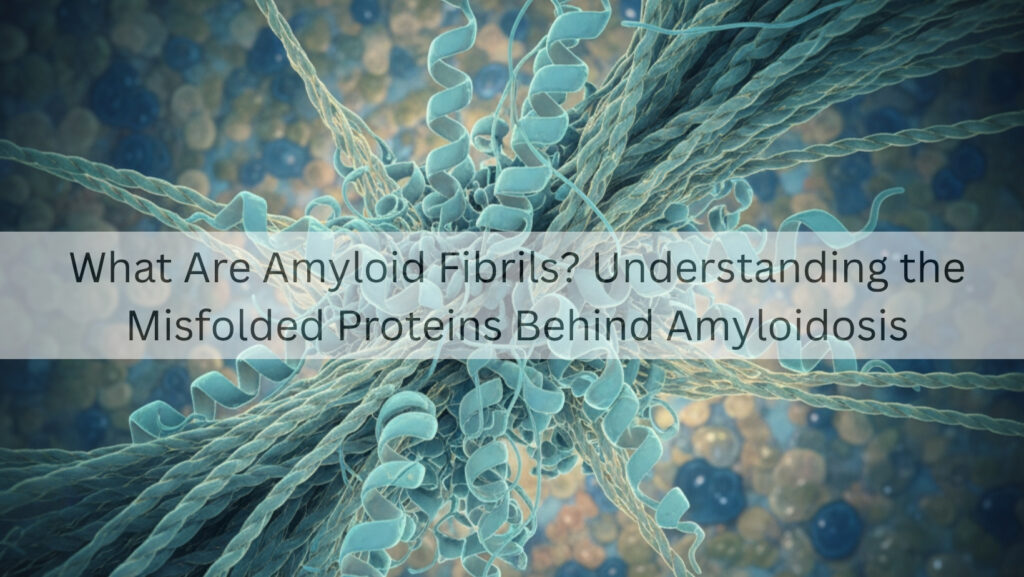
What Are Amyloid Fibrils?
Amyloid fibrils are long, rigid, insoluble protein fibers that form when misfolded proteins cluster together into structured, stick-like shapes. They have:
- Cross-beta sheet structure (a key feature of amyloid)
- Lengths from nanometers to micrometers
- High stability and resistance to breakdown
- The ability to attract and bind to other proteins and molecules
These fibrils accumulate in tissues and organs, creating what are known as amyloid deposits.
The Three Steps of Fibril Formation
- Misfolding
A soluble, normal protein takes on an abnormal shape. - Oligomerization
The misfolded proteins start sticking together, forming small clusters called oligomers. - Aggregation into Amyloid Fibrils
The oligomers bind together, elongate, and form fibrillar structures that deposit in tissues.
Why Does the Body Struggle to Clear Amyloid?
Amyloid fibrils are:
- Insoluble
- Highly stable
- Resistant to breakdown by enzymes
- Able to avoid the immune system
As these deposits grow, they crowd and harm tissues.
SECTION 3: Major Types of Amyloid Fibrils and Their Sources
Different diseases stem from different proteins that misfold.
1. AL Amyloid Fibrils
Formed from immunoglobulin light chains produced by abnormal plasma cells (similar to multiple myeloma). This is the most aggressive form.
2. ATTR Amyloid Fibrils
Produced from transthyretin (TTR) made in the liver. There are two types:
- Hereditary (mutant TTR)
- Wild-type (age-related)
3. AA Amyloid Fibrils
Formed from serum amyloid A, a protein that increases during inflammation.
4. Rare Types
Such as Aβ2M, ALECT2, AGel, AFib, etc.
Each type has its own specific clinical pattern.
SECTION 4: How Amyloid Fibrils Damage Organs
Amyloid fibrils harm the body through deposition and disruption.
1. Heart
Amyloid infiltrates the heart muscle, causing:
- Restrictive cardiomyopathy
- Diastolic dysfunction
- Arrhythmias
- Heart failure
- Conduction system disease
Heart involvement can be life-threatening.
2. Kidneys
Amyloid builds up in the glomeruli, leading to:
- Proteinuria
- Foamy urine
- Loss of kidney function
- Chronic kidney disease
3. Liver
Amyloid deposits result in:
- Hepatomegaly
- Congestive hepatopathy
- Increased liver enzyme levels
4. Gastrointestinal System
Amyloid impacts:
- Motility
- Nutrient absorption
- Autonomic function
5. Nervous System
Peripheral neuropathy and autonomic neuropathy are major issues in many forms, especially ATTR.
SECTION 5: The Cross-Beta Sheet Structure: The Signature of Amyloid
Amyloid fibrils are recognized by their cross-beta sheet arrangement. This structure gives them:
- Strength
- Stability
- Insolubility
- The ability to bind Congo red dye (orange-green birefringence)
Under electron microscopy, amyloid fibrils show:
- A non-branching structure
- A diameter of 7–13 nm
- A highly ordered arrangement
This is the gold standard for diagnosing amyloidosis.
SECTION 6: Why Amyloid Fibrils Are Toxic
Amyloid fibrils damage tissues through several mechanisms:
1. Space-Occupying Damage
As fibrils build up, they compress and push aside normal tissues.
2. Organ Stiffening
This is particularly problematic in the heart, where infiltration reduces elasticity.
3. Interference with Cell Function
Fibrils disrupt cell signaling, metabolism, and structure.
4. Toxic Oligomers
Before fibrils develop, smaller oligomers can be directly harmful to cells.
5. Interference with Blood Vessels
Amyloid build-up narrows and weakens blood vessels.
SECTION 7: Diagnosing Amyloid Fibrils in the Body
You can’t diagnose amyloid just with blood tests. Confirmation requires tissue biopsy and evidence of amyloid fibrils.
Diagnostic Methods
- Congo red staining (apple-green birefringence under polarized light)
- Electron microscopy
- Mass spectrometry for typing
- Immunohistochemistry
- Cardiac MRI (for heart involvement)
- Echocardiography
SECTION 8: Can Amyloid Fibrils Be Removed or Reversed?
The body can’t easily clear amyloid fibrils on its own. Treatment aims to:
- Stop further production of the precursor protein
- Remove existing deposits (newer therapies in ATTR)
- Support damaged organs
Treatments Vary by Type
- AL Amyloidosis: chemotherapy, stem cell transplant
- ATTR Amyloidosis: TTR stabilizers (tafamidis), silencers (patisiran, vutrisiran), gene therapies
- AA Amyloidosis: treat the underlying inflammation
SECTION 9: Why Understanding Amyloid Fibrils Helps Patients
Knowing what amyloid fibrils are helps patients:
- Understand their diagnosis
- Recognize severity
- Follow treatment
- Ask informed questions
- Make decisions about long-term care
This knowledge also helps caregivers offer better support.
Conclusion: Amyloid Fibrils Are the Center of Amyloidosis
Amyloid fibrils are not just simple protein deposits. They arise from significant molecular failures, biological stress, and abnormal protein behavior. They quietly invade organs, progress steadily, and need specialized treatment. Understanding fibrils is the first step to grasping amyloidosis itself.
The fight against amyloidosis starts with knowledge, and understanding amyloid fibrils is the bedrock of that understanding.
